3D Radiation Imaging for the Treatment of Advanced Stage Cancer
Treatment of advanced stage cancer requires appropriate therapy so as not to damage other healthy organs.
 whsiswanto
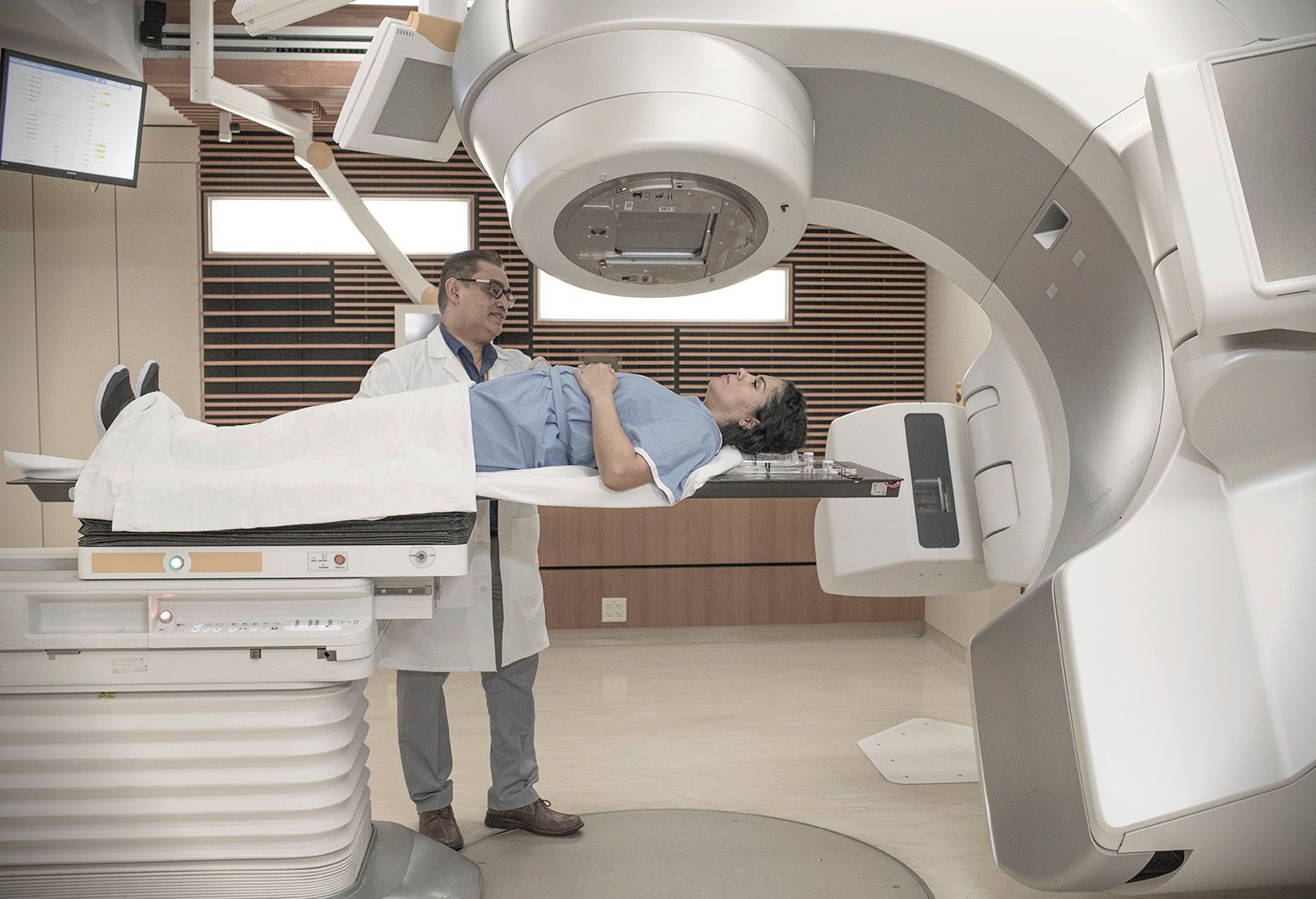
Radiation used to treat half of all cancer patients can now be measured during treatment for the first time with precise 3D imaging developed at the University of Michigan.
By capturing and amplifying the tiny sound waves created when X-rays heat tissue in the body, paramedics can map the dose of radiation in the body. This mapping process is able to provide doctors and other paramedics with the ability to guide treatment accurately and in real time regarding the condition of body tissues and the dosage required.
This is the first time this method has been used where interactions that previously could not be "seen" by doctors.
Once doctors start delivering radiation, the body becomes like a black box, as explained by Xueding Wang, the Jonathan Rubin Professor of Biomedical Engineering, professor of radiology and corresponding author of the study in Nature Biotechnology.
Initially, doctors don't know exactly where the X-rays hit inside the body and don't know how much radiation has been delivered to the target. And everybody is different, so making predictions for both aspects is tricky.
Radiation is used in treatment for hundreds of thousands of cancer patients each year, bombarding areas of the body with high-energy waves and particles, usually in the form of X-rays. The radiation can kill cancer cells directly or damage them so they cannot spread.
This benefit can damage other organs due to a lack of accuracy in the radiation dose and which tissues need to be X-rayed. This happens because radiation treatment often kills and damages healthy cells in the area around the tumour. It may also increase the risk of developing new cancers.
With real-time 3D imaging, doctors can more accurately direct radiation to cancer cells and limit exposure to adjacent tissue.
When X-rays are absorbed by tissues in the body, they are converted into heat energy. That heating causes the tissue to expand rapidly, and that expansion creates sound waves.
The acoustic waves are weak and usually undetectable by conventional ultrasound technology. Radiation acoustic imaging systems detect waves with an array of ultrasonic transducers placed at the patient's side. The signal is amplified and then transferred to an ultrasound device for image reconstruction.
With the images in hand, the oncology clinic can change the radiation levels or trajectory during the process to ensure safer and more effective treatment.
In the future, physicians may use imaging information to compensate for uncertainties arising from position, organ movement, and anatomical variations during radiation therapy. This imaging model will allow doctors to deliver doses to cancer tumours with pinpoint accuracy.
Another benefit of this technology is that it can be easily added to current radiation therapy equipment without drastically changing the processes doctors are used to.
In future applications, this technology could be used to personalize and adapt each radiation treatment to ensure normal tissue is kept at a safe dose and the tumour receives the desired dose. This technology would be particularly useful in situations where the target is in close proximity to radiation-sensitive organs such as the small intestine or stomach.
The University of Michigan has filed for patent protection and is looking for partners to help bring the technology to market. This research was supported by the National Cancer Institute and the Michigan Institute for Clinical and Health Research.
Image source: https://www.news-medical.net