Mouse Cortex Research for Treatment of Human Brain Cell Damage
The response of the human brain in the mouse cortex is the subject of research into treatments for brain cell damage in the future.

A team of engineers and neuroscientists has shown that human brain organoids implanted in mice have established functional connectivity to the animal's cortex and respond to external sensory stimuli. This research is good for treating damage to human brain cells due to stroke or other diseases in the future.
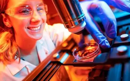
Human brain organoids inside the mouse cortex can react to visual stimuli in the same way as surrounding tissue, an observation the researchers were able to make in real time over several months thanks to an innovative experimental setup that combines transparent graphene microelectrode arrays and two-photon imaging.
The team led by Duygu Kuzum, a faculty member in the Department of Electrical and Computer Engineering at the University of California San Diego, detailed their findings in the December 26, 2022 issue of the journal Nature Communications. Kuzum's team collaborated with researchers from Anna Devor's lab at Boston University, Alysson R. Muotri's lab at UC San Diego, and Fred H. Gage's lab at the Salk Institute.
Human brain organoids in the mouse cortex originate from human-induced pluripotent stem cells, which are usually derived from skin cells. These brain organoids have recently emerged as promising models for studying human brain development, as well as various neurological conditions.
The researchers hope that this combination of innovative neural recording technology to study organoids will serve as a unique platform to comprehensively evaluate organoids as models for the treatment of brain cell damage and disease and investigate their use as neural prosthetics to restore the function of lost degenerated or damaged brain cells. Diseases such as stroke and brain cancer.
By placing an electrode array over the transplanted organoid, the researchers were able to electrically record neural activity from the implanted organoid and the surrounding host cortex in real-time. Using two-photon imaging, they also observed that the mice's blood vessels grew into organoids that provided the nutrients and oxygen needed for implants to treat damaged human brain cells.
But to date, no research team has been able to show that human brain organoids implanted in the cortex of mice are able to share the same functional properties and react to stimuli in the same way. This is because the technology used to record brain function is limited and generally unable to record activities that last only a few milliseconds.
The UC San Diego-led team was able to solve this problem by developing an experiment that combines microelectrode arrays made of transparent graphene and two-photon imaging, a microscope technique that can image living tissue up to one millimetre thick.

 whsiswanto